Last time: Spinodal decomposition--II.
Gradient energy
Elastic energy
Improved diffusion equation
Today: Spinodal decomposition--III; Nucleation--I.
Spinodal microstructures
Later stages
Nucleation theory: Homogeneous
The solution to the linearized diffusion equation that includes
gradient-energy effects has as its key feature an exponential growth
of composition waves over a range of wavenumbers ![]() inside the
coherent spinodal. One wavenumber,
inside the
coherent spinodal. One wavenumber, ![]() , receives the maximum
amplification. The theory predicts that the evolving microstructure
of growing waves will quickly become dominated by waves with
wavenmubers near
, receives the maximum
amplification. The theory predicts that the evolving microstructure
of growing waves will quickly become dominated by waves with
wavenmubers near ![]() . So one key aspect of spinodal
microstructures is spatial periodicity.
. So one key aspect of spinodal
microstructures is spatial periodicity.
In crystalline solutions, two additional points are important. First, at the early stages of decomposition, spinodal microstructures are always fully coherent and they can develop only within the coherent spinodal. Second, if the solution is elastically anisotropic, the wavevectors of the dominant waves will be aligned along elastically soft directions in the material. In cubic crystals, the <100> directions are usually the elastically soft directions .
Hence, in crystalline solutions, spinodal microstructures usually show both periodicity and alignment.
The kinetics of spinodal decomposition is usually studied by small-angle scattering techniques (light, x-rays, or neutrons). Spinodal microstructures in solids usually involve decomposition wavelengths on the order of 2-50 nm, so high-resolution imaging techniques like transmission electron microscopy or field-ion microscopy are employed.
The following images are typical of spinodal microstructures in metals and show examples of decomposition in elastically isotropic (Fe-Cr-Co) and anisotropic (Fe-Be) alloys imaged by field-ion microscopy. Images reproduced from S.S. Brenner, P.P. Camus, M.K. Miller, and W.A, Soffa, Acta metall. 32, 1217-1227 (1984).
 |
Later stages of spinodal decomposition
The linearized diffusion equation ignores several factors that limit
the validity of its solution for all but the shortest decompostion
times. One obvious deficiency is the assumption that
![]() is constant. This means that the decomposition can only be valid for
equal volume fractions of both phases, and also that the composition
amplitude will grow without limit. An additional freature missing in
the theory is interactions between growing waves, and these
interactions lead to gradual coarsening of the evolving two phase
structure. Most spinodal transformations observed experimentally show
increasing characteristic wavelengths with decomposition time, from
the earliest stages of the transformation. These nonlinear effects
are discussed in considerable detail in Cahn 1966.
is constant. This means that the decomposition can only be valid for
equal volume fractions of both phases, and also that the composition
amplitude will grow without limit. An additional freature missing in
the theory is interactions between growing waves, and these
interactions lead to gradual coarsening of the evolving two phase
structure. Most spinodal transformations observed experimentally show
increasing characteristic wavelengths with decomposition time, from
the earliest stages of the transformation. These nonlinear effects
are discussed in considerable detail in Cahn 1966.
Nucleation theory: Homogeneous
 |
Theory of steady-state nucleation
| (30-1) |
 |
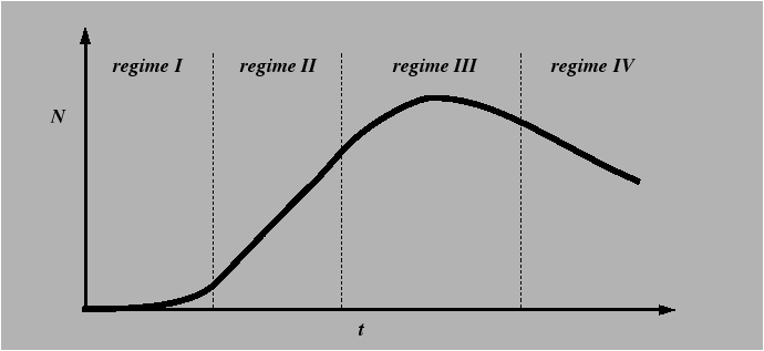
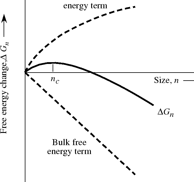
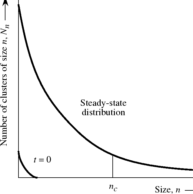
A theory for the steady-state nucleation regime must account for the
equilibrium distribution of clusters of size ![]() present in the
supersaturated solution. Fluxes in size-distribution space near the
critical size
present in the
supersaturated solution. Fluxes in size-distribution space near the
critical size ![]() can be used to develop a simple expression for
the steady-state nucleation rate.
can be used to develop a simple expression for
the steady-state nucleation rate.
Time-dependent nucleation
 |
Elastic strain energy effects on nucleation in solids
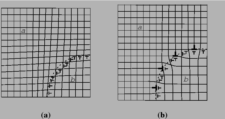
Interface structure, energy, and strain energy of inclusions
 |
Eshelby's method for calculating the elastic strain energy of coherent inclusions; some simple cases.