Computational Microscopy
Transmission electron microscopy works by accelerating electrons with a voltage difference and sending them towards a target. The electrons interact with the target and scatter. After the electrons have passed through the sample, they are focused with a magnetiic objective lens (or typically lenses). This lens produces a plane at which electrons scattered in the same direction arrive
at the same point---this is the diffraction pattern. Because diffraction transforms periodic elements into points, it is closely related to the fourier transform. An image of the target is created beyond this diffraction plane. An operator of an electron microscope can toggle between looking at the diffraction-plane or the image-plane.
Specific periodic features can be imaged by blocking all the electrons except those that appear at a point in the diffrations plance. To put it another way, one puts an aperature around a particular spot in order to selectively image the features that from that spot's periodicity. We can do this with a Fourier transform by selectling only a circular subset of points to be subsequently inversely transformed into an image.
The "Bright-Field Image" consists of using a central aperature around the direct beam to block off all others from contributing to the image.
The "Dark-Field Image" consists of selecting a specific diffracted peak with the aperature and using that to form an image
A "Structure image," or a "lattice image," uses the direct beam and one or more diffracted beams to form the image. In this case, the aperatures are typically much larger than for bright- or dark-field imaging.
Aperature size is effectively limited because of spherical aberrations that become significant for beams that are "off-axis" by a significant amount. So, in practice one can only use part of the Fourier spectrum (reciprocal space) to produce an image in TEM. You always lose some structure information in the image formation.
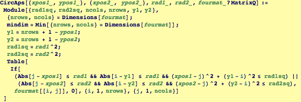
Function to Create Two Circular Aperatures (i.e, to remove all data from a Fourier Transform except the regions inside two specified circles):
CircAps[Center1, Center2, Radius1, Radius2, FourierData]

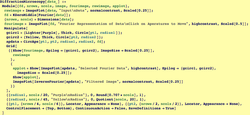
Function to Perform Diffraction Spot Microscopy on Real-Space Images
DiffractionMicroscopy[RealData]
Creates a Interactive Structure with Four Windows Arranged in a Square.
NorthWest: All Fourier Data from Real Image with Movable Aperatures, use a clicked mouse to move aperatures to various diffraction peaks.
NorthEast: The original real space image.
SouthWest: The Fourier image of the aperature filtered data. (Don't try to move these aperatures)
SouthEast: The reconstructed image from the aperature filtered data.

Function to Create Simulated Grain Structure for Subsequent Computational Microscopy Demo
GrainStructure[TotalSize, GrainSize]
Creates an array of "square grains" with stripes oriented in somewhat random orientations

| Created by Wolfram Mathematica 6.0 (01 November 2007) |